Brandon Snel1, Zachary Klukkert2 and Joydeep Chaudhuri1
1Central Michigan University College of Medicine
2Department of Anatomy and Cell Biology, Oklahoma State University Center for Health Sciences
*Correspondence to: Dr. Brandon Snel, Central Michigan University College of Medicine, USA
Received: Jan 20, 2025; Accepted: Jan 27, 2026; Published: Feb 11, 2025
Citation: Snel B (2025) Retroaortic Left Renal Vein in a Cadaver – Case Report. J Anatomical Variation and Clinical Case Report 3:13. DOI: https://doi.org/10.61309/javccr.1000
Copyright: ©2026 Snel B. This is an open-access article distributed under the terms of the Creative Commons Attribution License, which permits unrestricted use, distribution, and reproduction in any medium, provided the original author and source are credited.
ABSTRACT
In this case report, we aim to describe the local prevalence of retroaortic left renal vein (RLRV) in a university cadaver laboratory, were RLRV was defined as displacement of the left renal vein posterior to the abdominal aorta. Additionally, to explore the population prevalence of RLRV, its embryological origins, and recommendations for future identification and clinical significance. In our sample of 16 dissections, 1 (6.25%) were found to have RLRV. The population prevalence of RLRV relatively rare, with reports between 0.5-3.6%. Emryologically, at 8 weeks, a RLRV occurs if the intersubcardinal anastomosis improperly persists. Clinically, compression of the left rein vein can lead to hematuria, flank pain, and hypertension and ultimately posterior nutcracker syndrome. Identification can be achieved the adapter ultrasound and confirmatory contrast enhanced computer tomography. Retro aortic left renal vein is a relatively rare however clinically non-insignificant anatomical variation. Identification with appropriate imaging aid in surgical evaluations and diagnostic workups.
INTRODUCTION
Lower urinary tract symptoms such as urinary incontinence, bladder irregularity, voiding, and postmicturition are extremely common in adults aged 50 years and older, effecting over 40% of the world1. Hematuria, or the presentation of blood in urine, is one of the most common presenting symptoms in outpatient and emergency departments, therefore clinically significant sign that is both troubling to a patient and potentially indicative of several serious conditions2. The most common causes for hematuria are infection, glomerular disease, or trauma with a uncommon but nonsignificant cause being congenital renal vein anomalies. The gold standard for diagnosing hematuria is kidney biopsy, which can invasive and still lack definitive diagnosis. Unexplained, persistent hematuria can thus be the cause of some concern and may, in part, be explained by congenital anatomic variations in renal vasculature development.
RESULT
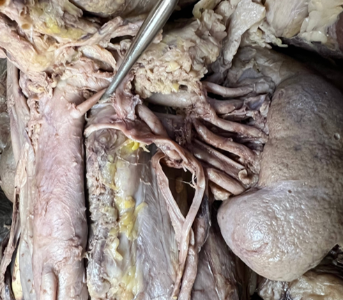
During a routine dissection of an 86 year old cadaver was found to have a retroaortic left renal vein (RLRV) with accessory vessels bilaterally. The ventral preaortic limb of the LRV is obliterated, but the dorsal retroaortic limb persists and joins the ICV in the orthotopic position. The left inferior phrenic, left gonadal vein, and left ureter are present and without significant variation. Distention of the ureter, hydronephrosis, and renal calculi were absent in both kidneys.
RLRV is defined as a displaced left renal vein (LRV) posterior to abdominal aorta and is relatively rare with reports between 0.5-3.6%3,4 in the general population. In our cadaveric sample of 16 dissections, 1 RLRV (6.25%) was found.
The cadaver was found to have a right-sided inferior vena cava (IVC) with the left renal vein projecting posteriorly to the abdominal aorta. Dual arterial supply to the left kidney, with one branch directly inferior to the superior mesenteric artery, and the second directly below with no distal branching pattern. On the right kidney, two veins connected the IVC, one midline to left vessels, and a second slightly posterior to origin of right gonadal vein (Figure 1).
DISCUSSION
Emryologically, at 8 weeks intersubcardinal anastomosis and intersupracardinal anastomosis form a collar of veins encircling the aorta. If the intersupracardinal anastomosis persists as the expected, an anterior aortic left renal vein develops. If the intersubcardinal anastomosis persists, a RLRV develops5.
Compression of the LRV can lead to posterior nutcracker syndrome, leading to blockage and dilatation of the left gonadal vein upstream of the inferior vena cava and varicocele of the gonadal veins6,7. Resulting symptoms include hematuria, flank pain, abdominal pain, and hypertension8.
Identification of RLRV is important surgically to prevent injury during renal transplantation, nephrectomy, repairing aortic aneurysms, and other retroperitoneal and abdominal surgery9. Doppler ultrasound and confirmatory contract-enhanced computed tomography can be used to identify RLRV10. The availability of point-of-care ultrasound and cost effectiveness makes it favorable.
RLRV is a rare but important anatomical variant to the venous system of the retroperitoneal space. Technology for identification is cheap, easy, and accessible and important in surgical procedures and explaining urological symptoms.
REFERENCES
1. Zhang AY, Xu X. Prevalence, Burden, and Treatment of Lower Urinary Tract Symptoms in Men Aged 50 and Older: A Systematic Review of the Literature. doi:10.1177/2377960818811773
2. Resorlu M, Sariyildirim A, Resorlu B, et al. Association of Congenital Left Renal Vein Anomalies and Unexplained Hematuria: Multidetector Computed Tomography Findings. Urol Int. 2015;94(2):177-180. doi:10.1159/000365664
3. Heidler S, Hruby S, Schwarz S, Sellner-Zwieauer Y, Hoeltl W, Albrecht W. Prevalence and incidence of clinical symptoms of the retroaortic left renal vein. Urol Int. 2015;94(2):173-176. doi:10.1159/000367697
4. Karaman B, Koplay M, Özturk E, et al. Retroaortic left renal vein: Multidetector computed tomography angiography findings and its clinical importance. Acta radiol. 2007;48(3):355-360. doi:10.1080/02841850701244755
5. Kyung D-S, Lee J-H, Shin D-Y, Kim D-K, Choi I-J. The double retro-aortic left renal vein. Anat Cell Biol. 2012;45(4):282. doi:10.5115/acb.2012.45.4.282
6. Almuqamam M, Ebrahim M, Nassar G, Kaplan M. Atypical Posterior Nutcracker Syndrome in a 17-Year-Old Male Without Hematuria. Cureus. 2021;13(8). doi:10.7759/CUREUS.17221
7. Lizama L, Bran S, Kuestermann S, García-Gallont R. RENAL VEIN THROMBOSIS DUE TO POSTERIOR NUTCRACKER SYNDROME. Methodist Debakey Cardiovasc J. 2020;16(4). doi:10.14797/MDCJ-16-4-E5
8. Nam JK, Park SW, Lee SD, Chung MK. The clinical significance of a retroaortic left renal vein. Korean J Urol. 2010;51(4):276-280. doi:10.4111/KJU.2010.51.4.276
9. Karkos CD, Bruce IA, Thomson GJL, Lambert ME. Retroaortic left renal vein and its implications in abdominal aortic surgery. Ann Vasc Surg. 2001;15(6):703-708. doi:10.1007/s10016-001-0022-y
10. Rampersad F, Chan A, Diljohn J. Retroaortic left renal vein (RLRV) draining into the left common iliac vein: A rare variant and its clinical implication. BMJ Case Rep. 2019;12(5):1-2. doi:10.1136/bcr-2019-230004