Anthony Mufarreh1*, Brandon Snel1, Zachary Klukkert2 and Joydeep Chaudhuri1
1Central Michigan University College of Medicine
2Department of Anatomy and Cell Biology, Oklahoma State University Center for Health Sciences
ABSTRACT
In this case report, we aim to describe the local prevalence of retroaortic left renal vein (RLRV) in a university cadaver laboratory, were RLRV was defined as displacement of the left renal vein posterior to the abdominal aorta. Additionally, to explore the population prevalence of RLRV, its embryological origins, and recommendations for future identification and clinical significance. In our sample of 16 dissections, 1 (6.25%) were found to have RLRV. The population prevalence of RLRV relatively rare, with reports between 0.5-3.6%. Emryologically, at 8 weeks, a RLRV occurs if the intersubcardinal anastomosis improperly persists. Clinically, compression of the left rein vein can lead to hematuria, flank pain, and hypertension and ultimately posterior nutcracker syndrome. Identification can be achieved the adapter ultrasound and confirmatory contrast enhanced computer tomography. Retro aortic left renal vein is a relatively rare however clinically non-insignificant anatomical variation. Identification with appropriate imaging aid in surgical evaluations and diagnostic workups.
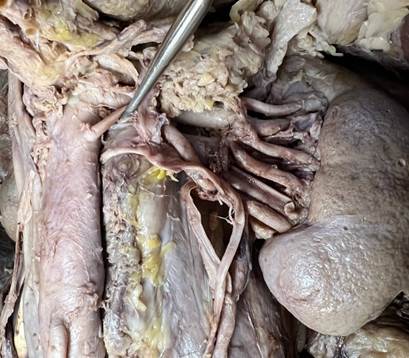
Figure 1: Anterior-posterior view of retroperitoneal area at level of the kidney. Left kidney shown. RLRV circled in red. Left renal artery indicated by probe tool.