Mehri Fayazi¹*, Blanca Castillo¹, Noura Daryani¹, Yuan Hu¹, Kristin Devaney¹, Ravi Lai¹ and Aliasghar Arabi Mianroodi2
¹Woody L. Hunt School of Dental Medicine, Texas Tech University Health Sciences Center El Paso, El Paso, TX, USA
²Paul L. Foster School of Medicine, Texas Tech University Health Sciences Center El Paso, El Paso, TX, USA
*Correspondence to: Dr. Mehri Fayazi, Assistant Professor, Woody L. Hunt School of Dental Medicine, Texas Tech University Health, Sciences Center El Paso, El Paso, TX, USA
Received: Aug 31, 2026; Accepted: Sep 14, 2026; Published: Sep 21, 2026
Citation: Fayazi M, Castillo B, Daryani N, Hu Y, Devaney K, et al. (2026) Complete Ureteral Duplication and an Accessory Renal Artery: Two Case Reports. J Anatomical Variation and Clinical Case Report 4:127. DOI: https://doi.org/10.61309/javccr.1000127
Copyright: ©2026 Fayazi M. This is an open-access article distributed under the terms of the Creative Commons Attribution License, which permits unrestricted use, distribution, and reproduction in any medium, provided the original author and source are credited.
ABSTRACT
Background: Anatomical variations of the urinary collecting system and renal vasculature are clinically important because they may affect diagnostic imaging as well as surgical and interventional procedures involving the kidneys, ureters, and retroperitoneum. These case reports describe two developmental variations identified during routine human donor dissection: complete unilateral ureteral duplication and an accessory renal artery (ARA). Methods: As part of the Distinction in Dental Human Anatomy (DDHA) program, dental students performed routine dissection of the abdomen. The abdominal and retroperitoneal structures were carefully dissected to expose the kidneys, renal vessels, renal pelvises, and ureters while preserving their anatomical relationships. We traced the ureters from the renal pelvises toward the urinary bladder, and traced the renal arteries from the abdominal aorta to the kidneys. We carefully dissected and photographically documented the identified variations. The study protocol was reviewed and approved by the Institutional Biosafety Committee (IBC #26006). Results: We identified two anatomical variations in separate human donors. A 97-year-old female donor demonstrated complete unilateral duplication of the right ureter, with two distinct renal pelvises each giving rise to a separate ureter that remained distinct throughout its course to the urinary bladder. A 76-year-old male donor demonstrated a left ARA arising independently from the lateral aspect of the abdominal aorta, inferior to the main left renal artery. The accessory artery was smaller in caliber than the main renal artery and coursed directly to the inferior pole of the left kidney rather than entering through the renal hilum. Conclusion: Complete ureteral duplication and ARA represent clinically relevant developmental variations that may influence urologic, vascular, transplant, and retroperitoneal procedures. Recognition of these variations is important for accurate interpretation of diagnostic imaging and prevention of inadvertent vascular or ureteral injury during surgical procedures. These findings also highlight the value of human donor dissection for understanding anatomical variability and integrating gross anatomy, embryology, and clinical practice.
Keywords: Accessory renal artery (ARA); Duplicated renal pelvis; Ureteral duplication
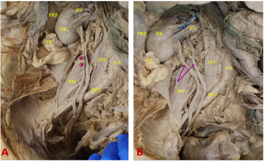
Figure 1A and 1B: Abdominal dissection following removal of the pararenal fat, renal fascia (Gerota’s fascia), and perirenal fat, exposing the kidneys and ureters. Complete unilateral duplication of the right ureter is demonstrated, with two distinct renal pelvises giving rise to two separate ureters. The duplicated right ureters were traced inferiorly across the pelvic brim and remained separate throughout their course to the urinary bladder. Two pink pins and pink arrows indicate the duplicated right ureters. Abbreviations: AA: abdominal aorta; IVC: inferior vena cava; RK: right kidney; RCI: right common iliac artery; PM: right psoas major muscle; PF: perirenal fat; PRF: prerenal fascia; RV: right renal vein; and blue arrow: right renal artery.