*Correspondence to: Dr. Macario Llamas, Associate Professor, Anatomy and Clinical Medicine, Kirk Kerkorian School of Medicine at UNLV, USA
Received: May 30, 2025; Accepted: Jun 12, 2025; Published: Jun 20, 2025
Citation: Alkhouri S, Asefirad A, Bakalov Y, Joshi A, Khorsandi J, et al. (2025) Multifocal Thymic Tumors with Systemic Hypercoagulability: A Rare Autopsy Case Report. J Anatomical Variation and Clinical Case Report 3:119. DOI: https://doi.org/10.61309/javccr.1000119
Copyright: ©2025 Alkhouri S. This is an open-access article distributed under the terms of the Creative Commons Attribution License, which permits unrestricted use, distribution, and reproduction in any medium, provided the original author and source are credited.
ABSTRACT
Thymic tumors (TM) are rare epithelial neoplasms that are typically associated with autoimmune disorders, but their potential role in systemic hypercoagulability remains largely unexplored. We report an unusual autopsy case of a deceased male in his 60s, in which seven distinct TM were identified throughout his mediastinum. Histologic analysis confirmed WHO subtype B2 thymomas, characterized by lobulated architecture, abundant lymphocytes, and focal necrosis. Strikingly, multiple ante-mortem thrombi were found in the coronary, pulmonary, mesenteric, and carotid arteries, raising concern for a paraneoplastic hypercoagulable state akin to Trousseau’s syndrome. While thrombotic complications are well-recognized in other malignancies, they are exceedingly rare in thymomas, particularly in cases with multifocal involvement and widespread arterial thrombosis. This case highlights the need for increased clinical awareness of potential coagulation abnormalities in thymic neoplasms and supports further investigation into their pathophysiological mechanisms.
Keywords: Thymic tumors; Multifocal thymomas; Hypercoagulability; Paraneoplastic thrombosis; Arterial thromboembolism; Systemic coagulopathy; Mediastinal neoplasms
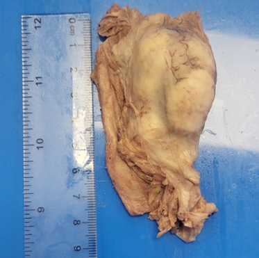
Figure 1: Gross pathology of a resected thymoma from the mediastinum. The mass is well-circumscribed and encapsulated, displaying a tan-white lobulated cut surface with focal cystic changes.