Varuneshwar Parsad1* and Mamata Srinivasan2
1Department of Human Body Structure and Function, Medical University of the Americas, Saint Kitts and Nevis, West Indies
2Department of Human Histology and Physiology, Medical University of the Americas, Saint Kitts and Nevis, West Indies
*Correspondence to: Dr. Varuneshwar Parsad, Human Body Structure and Function, Medical University of the Americas, Saint Kitts and Nevis, West Indies
Received: Jan 08, 2025; Accepted: Jan 30, 2025; Published: Feb 05, 2025
Citation: Parsad V, Srinivasan M (2025) An Atypical Branch from the Right Pulmonary Artery During Heart Dissection: A Case Report. J Anatomical Variation and Clinical Case Report 3:115. DOI: https://doi.org/10.61309/javccr.1000115
Copyright: ©2025 Parsad V. This is an open-access article distributed under the terms of the Creative Commons Attribution License, which permits unrestricted use, distribution, and reproduction in any medium, provided the original author and source are credited.
ABSTRACT
Anatomical variations in the pulmonary vasculature are rare but clinically significant, particularly in surgical and interventional cardiology. During a routine heart dissection of a 75-year-old female donor at the Medical University of the Americas, an atypical branch originating from the right pulmonary artery was observed. This branch, measuring approximately 5 mm in diameter, coexisted with an otherwise normal right pulmonary artery. Pulmonary artery anomalies stem from complex embryological processes and have been linked to conditions such as pulmonary artery slings and Kabuki syndrome. Recognizing these variations is crucial for accurate diagnostic imaging, surgical planning, and interventional procedures to prevent complications. This case contributes to the growing body of literature on pulmonary vascular anomalies and highlights the importance of meticulous anatomical evaluation in clinical practice.
Keywords: Right pulmonary artery; Anatomical variation; Pulmonary artery anomaly; Cardiovascular anatomy; Heart dissection; Atypical vascular branch
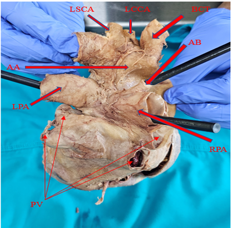
Figure 1: (Posterior surface or base of heart) AA (arch of aorta), BCT (brachiocephalic trunk), LCCA (left common carotid artery), LSCA (left subclavian artery), AB (atypical branch), RPA (right pulmonary artery), LPA (left pulmonary artery), PV (pulmonary veins).