*Correspondence to: Rozi Mahmud, Department of Veterinary Preclinical Sciences, Faculty of Veterinary Medicine, University Putra Malaysia, 43400 UPM, Serdang Malaysia
Received: Sep 28, 2020; Accepted: Oct 15, 2020; Published: Oct 20, 2020
Citation: Mahmud R (2020) Biocompatibility Assessment and Cellular Uptake of Conjugated Gold-Cockle Shell Derived Calcium Carbonate Nanoparticles for Fluorescent Imaging. J Nanomed Nanotech Nanomat. 1: 104.
Copyright: ©2020 Mahmud R, et al. This is an open-access article distributed under the terms of the Creative Commons Attribution License, which permits unrestricted use, distribution, and reproduction in any medium, provided the original author and source are credited.
Abstract
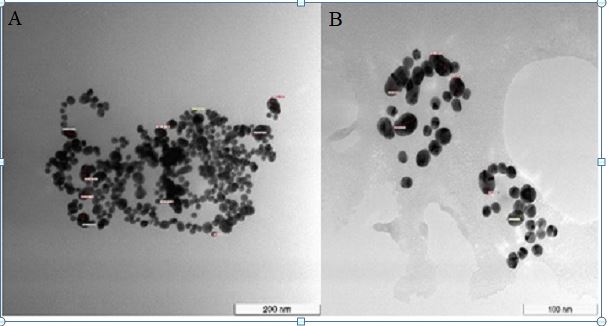
In recent years, fluorescent imaging (FI) has emerged as a major area of interest within the field of medical imaging. FI plays a critical role in molecular imaging. There is evidence suggesting its utility in providing a detailed view of biological and cellular processes at a molecular level. This study aimed to assess the biocompatibility and cellular uptake of conjugated gold-cockle shell derived calcium carbonate nanoparticles (Au-CsCaCO3NPs) for fluorescent imaging. Researchers have shown a keen interest in the development of targeted multifunctional agents in oncology and near infrared (NIR) fluorescence imaging. This is expected to have significant impact on medical imaging due to the low tissue auto fluorescence and high cellular penetration within the NIR spectrum window. Imaging agents are known to be associated with risks such as non-biodegradability and high toxicity. The synthesized Au-CsCaCO3NPs were characterized for size and morphology, zeta potential and absorbance in the UV-Vis spectrum. Biocompatibility of Au-CsCaCO3NPs in cultured human breast carcinoma cells (MCF-7) and mouse embryonic fibroblast cells (NIH-3T3) was evaluated using lactate dehydrogenase (LDH) and reactive oxygen species (ROS) bioassays for toxicity analysis. Cellular morphology and uptake was examined by fluorescence and confocal microscopy. Cells were able to take up nanoparticles within their cellular compartments. Further, increased cell death was observed in Au-CsCaCO3NPs-treated MCF-7 cells relative to Au-CsCaCO3NPs-treated NIH-3T3 cells. The Au-CsCaCO3NPs were biocompatible, environmentally friendly and easily synthesized. These results suggest Au-CsCaCO3NPs may have significant cellular imaging utility.
Keywords: Au-CsCaCO3NPs; Nanoparticle biocompatibility; LDH; ROS; Cellular uptake; Fluorescent imaging